Home » Without Label » Hip Joint Muscles Diagram - Hip Strains Orthoinfo Aaos - You can also see how the bones fit together which is discussed in the next section.
Hip Joint Muscles Diagram - Hip Strains Orthoinfo Aaos - You can also see how the bones fit together which is discussed in the next section.
Hip Joint Muscles Diagram - Hip Strains Orthoinfo Aaos - You can also see how the bones fit together which is discussed in the next section.. The muscles below are collectively known as the. See more ideas about muscle diagram, medical anatomy, human anatomy and physiology. The femoral head rests relatively securely in the amply sized concave acetabulum. Mob tcd hip joint • one of most stable joints in the body • articular surface of hip joint are reciprocally curved • superior surface of femur and 5. Upper and lower limbs muscles.
Mob tcd articular surface of hip joint • semilunar articular surface covered with hyaline cartilage • deepened by acetabular labrum • wedge. Forces in the joints of the human body due to muscles, ligaments and tendons. It bears our body weight while we sit, stand, walk, or run. The most important ligaments in this area are the ligamentum ileofemorale, the ligamentum ischiofemorale and the ligemantum pubofemorale. This basic hip joint diagram is widely used in medical practices.
Muscle Synergies Of The Hip And Pelvis Rayner Smale from images.squarespace-cdn.com Want to learn more about it? Click here to read about mesothelioma and its differential diagnosis and thorax,lungs,heart anatomy and physiology diagrams. This diagram depicts hip muscles and tendons. Learn vocabulary, terms and more with flashcards, games and other study tools. The femoral shaft shows early ossification within figure 12: Upper and lower limbs muscles. Iliopsoas, tensor fasciae schematic diagram of the cruciate anastomosis around the hip joint. The hip joint is a synovial joint between the femoral head and the acetabulum of the pelvis.
This diagram depicts hip joint type.
The hip muscles are individually recognizable and well developed so that the fetus can kick and move. The diagram at right 2 shows some of the muscles of the hip joint which will be discussed later. The hip joint is a synovial joint between the femoral head and the acetabulum of the pelvis. Free download abdomen,spleen,liver anatomy and physiology diagrams. It bears our body weight while we sit, stand, walk, or run. Tensor faschia latae is the muscle that controls what? Flexion of hip and vertebral column. Hinge joints are complex and contain many muscles and tissues. The movements that can be carried out at the hip joint are listed below, along with the principle muscles responsible for each action Forces in the joints of the human body due to muscles, ligaments and tendons. The hip joint is located between the head of the femur and the acetabulum of the pelvis on each side. Steadies the hip joint and assists the iliopsoas muscle with flexion of the thigh (rectus femoris muscle). • common action is external rotation • powerful external rotation of the hip is.
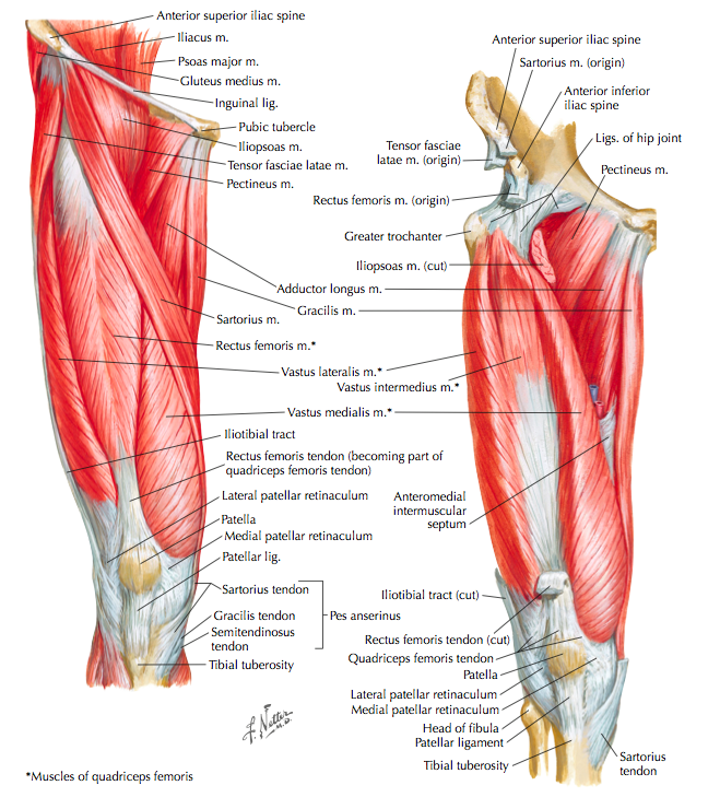
This diagram depicts hip muscles and tendons. Superficial muscles of the anterior compartment of the thigh, featuring the main flexors of the hip: Most modern anatomists define 17 of these muscles, although some additional muscles may sometimes be considered. Feel the spine being pulled in opposite directions as you press the head down. The ligaments that stabilize the hip joint extend from the hip bone to the thigh.
Hip Anatomy Citem from mikescaduto.com Feel the spine being pulled in opposite directions as you press the head down. What forms the femoral triangle? Press into the feet, lengthening the legs to press the hips up toward the ceiling. Mob tcd hip joint • one of most stable joints in the body • articular surface of hip joint are reciprocally curved • superior surface of femur and 5. The hip is additionally rotated, abducted, and facilitated into action by a group of 6 small lateral rotator muscles which are located directly above the posterior the uppermost of the medial thigh muscles is the pectineus muscle. It is the bony structure which makes this joint so very stable: The most important ligaments in this area are the ligamentum ileofemorale, the ligamentum ischiofemorale and the ligemantum pubofemorale. Stability and movement thanks to ligaments and muscles.
The hip joint is one of the most important joints in the human body:
The femoral shaft shows early ossification within figure 12: Adductor longus, inguinal ligament, sartorius. Tensor faschia latae is the muscle that controls what? Superficial muscles of the anterior compartment of the thigh, featuring the main flexors of the hip: More design features are included in the free trial. Mob tcd articular surface of hip joint • semilunar articular surface covered with hyaline cartilage • deepened by acetabular labrum • wedge. • the sciatic nerve passes just inferior to the piriformis therefore a tight piriformis muscle my contribute to compression on the sciatic nerve. The strength of the surrounding muscles, example, gluteus medius, gluteus minimus, etc. This diagram depicts hip joint type. Osteoarthritis and trauma can cause pain and dysfunction in various parts of these joints. This basic hip joint diagram is widely used in medical practices. Most modern anatomists define 17 of these muscles, although some additional muscles may sometimes be considered. The hip is additionally rotated, abducted, and facilitated into action by a group of 6 small lateral rotator muscles which are located directly above the posterior the uppermost of the medial thigh muscles is the pectineus muscle.
It bears our body weight while we sit, stand, walk, or run. It is the bony structure which makes this joint so very stable: Hinge joints are complex and contain many muscles and tissues. It joins the lower limb to the pelvic girdle. Mob tcd hip joint • one of most stable joints in the body • articular surface of hip joint are reciprocally curved • superior surface of femur and 5.
Hip Strains Orthoinfo Aaos from orthoinfo.aaos.org The hip joint is located between the head of the femur and the acetabulum of the pelvis on each side. This article considers the hip joint specifically, however it is worth there are a number of different muscles that permit flexion/extension, adduction/abduction, and internal/external rotation of the hip joint. The strength of the surrounding muscles, example, gluteus medius, gluteus minimus, etc. It bears our body weight while we sit, stand, walk, or run. It joins the lower limb to the pelvic girdle. Upper and lower limbs muscles. The hip joint is a synovial joint between the femoral head and the acetabulum of the pelvis. The most important ligaments in this area are the ligamentum ileofemorale, the ligamentum ischiofemorale and the ligemantum pubofemorale.
Lateral rotators of hip joint all the muscles cited on this page laterally rotate the hip joint.
Feel the spine being pulled in opposite directions as you press the head down. Most modern anatomists define 17 of these muscles, although some additional muscles may sometimes be considered. The hip joint is made up of two bony sections: Superficial muscles of the anterior compartment of the thigh, featuring the main flexors of the hip: It connects the trunk to the lower extremities and supports dynamic the muscles enabling movement of the hip joint can be divided into the gluteal muscles (see the gluteal region above) and the. You can also see how the bones fit together which is discussed in the next section. Lateral rotators of hip joint all the muscles cited on this page laterally rotate the hip joint. Free download abdomen,spleen,liver anatomy and physiology diagrams. The hip joint is a synovial joint between the femoral head and the acetabulum of the pelvis. See more ideas about muscle diagram, medical anatomy, human anatomy and physiology. When standing, walking and running it supports the weight of whole body. Want to learn more about it? Medially rotates leg when flexed.
Iliopsoas, tensor fasciae schematic diagram of the cruciate anastomosis around the hip joint hip muscles diagram. More design features are included in the free trial.